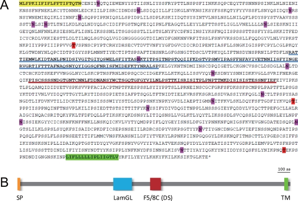
Fig 2.
Expected structure of MacA. (A) The deduced amino acid sequence is shown. The yellow and green highlights indicate the signal peptide (SP) and transmembrane (TM) sequences, respectively. Possible N-glycosylation (pink) and O-glycosylation (red) sites are also highlighted. Blue and red underlines represent LamGL and F5/8C (discoidin [DS]) domains, respectively. O-glycosylation sites were predicted using the NetOGlyc 3.1 server (http://www.cbs.dtu.dk/services/netoglyc/). (B) The overall structure of MacA is shown schematically.

