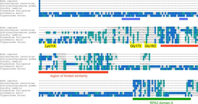
Fig 1.
Multiple-sequence alignment of the RPA2 N terminus. A multiple-sequence alignment was generated from a data set of 16 eukaryotic RPA2 proteins, 7 of which were selected for display in this figure. Residues are shown with a blue or cyan background indicating either identity or similarity to the majority consensus, respectively. Yellow flags on the T. brucei sequence indicate the residues tagged in this study. Blue bars mark stretches rich in basic residues in the trypanosomatid homologues. The red bar marks the region of limited similarity; the green bar marks RPA2 domain A.

