Fig 2.
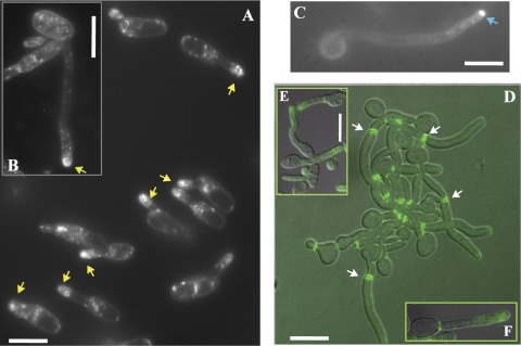
Shmooing (A, B, E, and F) and hyphal (C and D) cells of C. albicans. (A to C) Cells were stained with FM4-64. In shmooing opaque cells obtained from a mating mixture (MTLa/MTLa NOP1/NOP1-YFP MYO5/MYO5-GFP and MTLα/MTLα MYO5/MYO5-GFP strains) after 5 to7 h (A) and 16 h (B) of cultivation in liquid Spider medium at 24°C, we observed a diffuse bright spot (yellow arrows) at the tip of the mating projection. In hyphal cells (C) generated by overnight growth in YPD medium at 30°C, followed by dilution to an OD600 of 0.15 and cultivation in YPD medium supplemented with 10% (wt/vol) fetal calf serum at 37°C for 2, h we observed a tight fluorescent spot characteristic of a classic Spitzenkörper (blue arrow). (D to F) CDC12/CDC12-GFP cells were examined for Cdc12-GFP staining. Cdc-12p tagged with GFP localizes at the neck as bars parallel to the projection axis in both hyphal cells (D) and shmoos (E); however, although septin rings are evident in germ tubes (white arrows) (D), we never see similar structures inside mating projections (E and F). Fluorescent images of Cdc12p are superimposed on differential interference contrast images (D and E). Bars, 3 μm (A, B, C, D, and F) and 5 μm (E).

