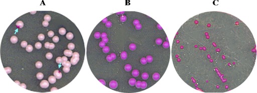
Fig 4.
Identification of opaque-phase cells on SCD plates containing phloxine B. (A) MTLa/MTLa MYO5/Δmyo5 cells. Arrows point to colonies with sectors containing opaque cells. (B) MTLa/MTLa Δ/Δ myo5 cells. (C) MTLa/MTLa MET3-WOR 1 Δ/Δ myo5 cells grown on activating SCD-MC medium to induce WOR1 expression. This treatment generates colonies that are dark pink and smaller than MTLa/MTLa Δ/Δ myo5 and MTLa/MTLa MYO5/Δmyo5 colonies. Cells in these colonies have an abnormal morphology.

