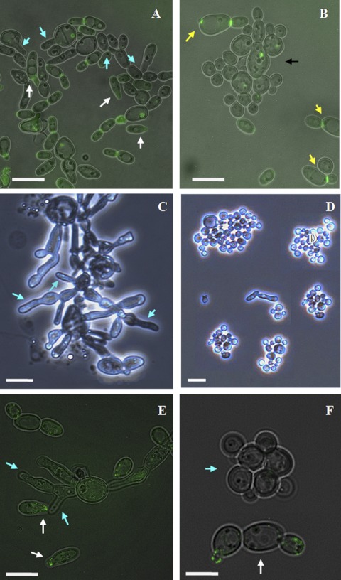
Fig 9.
Polarized growth in strains with conditional and true knockouts of the MYO5 gene. Mating mixtures consist of MTLa/MTLa MET3-MYO5/Δmyo5 cells from “wrinkled” or “smooth” colonies and MTLα/MTLα CDC12/CDC12-GFP opaque cells (A and B) or a/a MET3-WOR1 Δ/Δ myo5 and MTLα/MTLα MYO5/MYO5-GFP cells (E and F) (see the explanation in the text). (A) Cells from “wrinkled” colonies are still able to generate shmoos (blue arrows) and stimulate shmooing in α/α opaque cells with the CDC12-GFP marker (white arrows). (B) Cells from “smooth” colonies (black arrow) demonstrate white morphology and are unable to shmoo. Yellow arrows point to opaque MTLα/MTLα CDC12/CDC12-GFP cells that also do not shmoo. (C and D) Phenotypes of pure a/a MET3-WOR1 Δ/Δ myo5 cells in initial cultures in SCD-MC and SCD+MC liquid media, respectively. Blue arrows show shmoo-like formation in cells grown under MET3-inducing conditions (C). (E) Δ/Δ myo5 cells with ectopic expression of WOR1 generate the same shmoo-like formations (blue arrows) in the mating mixture as in the initial culture. White arrows show shmoos in opaque α/α cells with the MYO5-GFP marker. (F) Under WOR1 shutoff conditions. MTLa/MTLa MET3-WOR1 Δ/Δ myo5 cells (blue arrow) convert to white phase, and the opaque α/α MYO5/MYO5-GFP cells (white arrow) do not form shmoos. Fluorescent images are superimposed on differential interference contrast images. Bars, 5 μm (A, E, and F), 4 μm (B), 10 μm (C), and 12.7 μm (D). An inverted microscope was used. Magnification, ×40.

