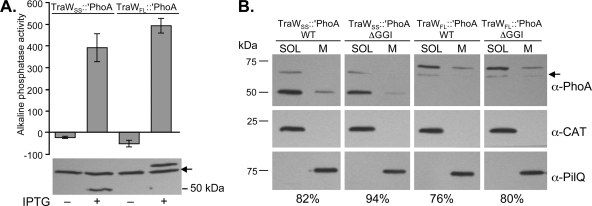
Fig 7.
Activity and localization of TraW::PhoA fusions in N. gonorrhoeae. (A) Alkaline phosphatase activity and immunoblot analysis of N. gonorrhoeae strains producing TraWSS::′PhoA (MR544) and TraWFL::′PhoA (MR553). Alkaline phosphatase units were normalized to total mg of protein. The mean ± standard deviation of three independent experiments is shown. Arrow, a band that cross-reacts with the anti-PhoA antibody. (B) Subcellular localization of TraW::PhoA fusions in gonococcal cells. Four chloramphenicol-resistant strains were constructed: MR555 (expressing traWSS::′phoA in a WT background), MR561 (expressing traWSS::′phoA in a ΔGGI background), MR556 (expressing traWFL::′phoA in a WT background), and MR562 (expressing traWFL::′phoA in a ΔGGI background). Cells were fractionated into a soluble fraction (SOL) containing the cytoplasm and periplasm as well as a membrane fraction (M) containing the inner and outer membranes. Subcellular fractions were probed with antibodies against alkaline phosphatase (PhoA), the cytoplasmic CAT, and the outer membrane protein PilQ. Densitometry analysis was performed using ImageJ software. The percentage of total TraW::PhoA fusion protein associated with the soluble fraction for each strain is reported. Arrow, a band that cross-reacts with the anti-PhoA (α-PhoA) antibody.

