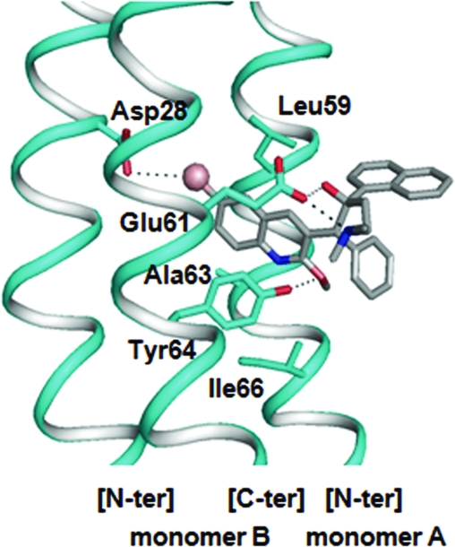
Fig 4.
Optimal docking result obtained for the R,S stereoisomer of TMC207 in the C ring of M. tuberculosis. The polypeptide chains forming the binding cleft are shown in ribbon representation. The amino acid residues contributing to the binding region are shown in stick representation. Oxygen and nitrogen atoms are indicated in red and dark blue, respectively. The TMC207 molecule is represented by using CPK colors, and the bromine atom is shown as a sphere. Hydrogen and halogen bonds are represented by dotted lines.

