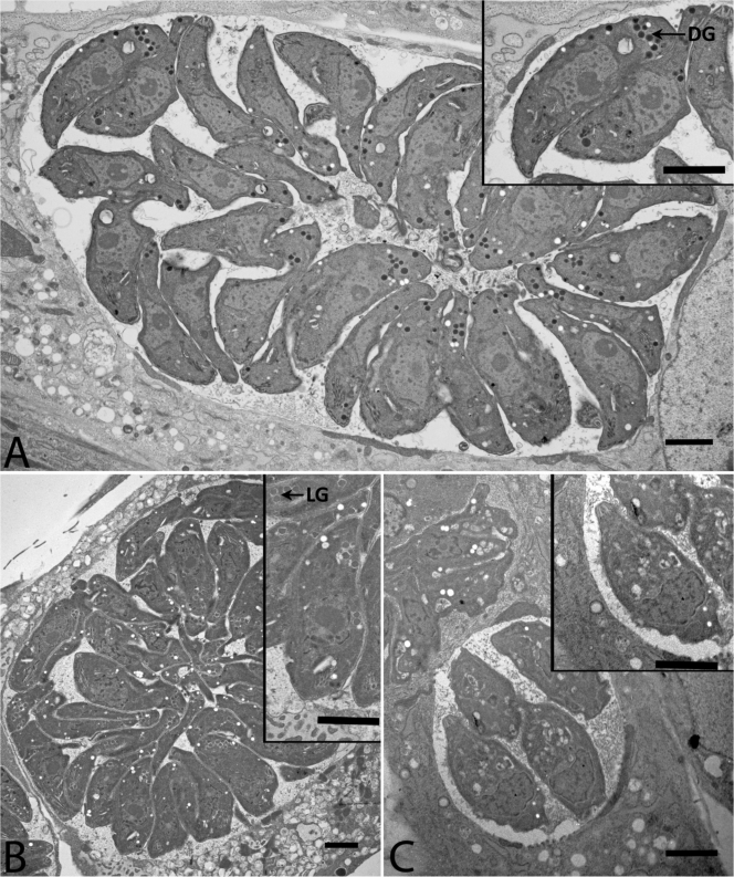
Fig 3.
Electron microscopy images showing loss of dense granule content and acidocalcisomes after treatment with imides. (A) Wild-type, untreated RH tachyzoite parasites. (Inset) Enlargement. Note normal ultrastructure. DG, dense granules. (B) Tachyzoites treated with MP-IV-1. (C) Tachyzoites treated with QQ-437. Note the absence of dense granules with normal ultrastructure and density in N-benzoyl-2-hydroxybenzamide-treated parasites. LG, light granules.

