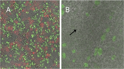
Fig 6.
Combined fluorescence and light microscopic investigations at day 4 of biofilms of P. aeruginosa exposed to PMNs (arrow) for 180 min at 37°C and then subsequently stained with the DNA stain PI. (A) Biofilm grown without ajoene in the medium; (B) biofilm grown in the presence of 100 μg/ml ajoene in the medium. Red fluorescence indicates lysed PMNs, and green fluorescence indicates the P. aeruginosa biofilm.

