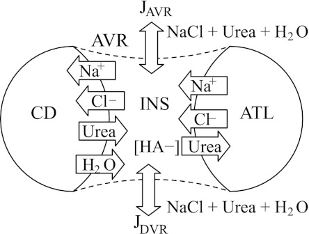
Fig. 2.
Schematic diagram of compartment model. Represented are cross-sections of CD, ascending thin limb (ATL), and AVR, which interact through the interstitial nodal space (INS). Fractions of the CD and ATL are assumed to be in direct contact with the AVR endothelium. Cross-section of a DVR is also represented to model limited interactions between intra- and extra-cluster structures. JAVR and JDVR, transmural water flux in AVR and DVR; HA, hyaluron.

