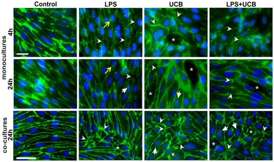
Figure 7. Lipopolysaccharide (LPS) and unconjugated bilirubin (UCB) modify the distribution of β-catenin in brain endothelial cells.
Cells in mono-culture or co-cultured with astrocytes were fixed and immunostained with an antibody against β-catenin to evaluate its cellular localization (scale bars, 40 and 20 µm, respectively). Disruption of the monolayer with gaps between endothelial cells (*), alterations in protein patterns (arrowheads) with the presence of dot-like staining (yellow arrow), and perinuclear distribution (arrows) are indicated. Representative results from one of two independent experiments are shown.

