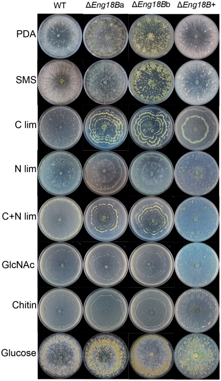
Figure 3. Colony morphology of WT, ΔEng18B and ΔEng18B+ T. atroviride strains in different nutrient regimes.
T. atroviride strains were inoculated on solid PDA, SMS, C limitation, N limitation, C+N limitation, N-acetylglucosamine (GlcNAc), chitin and glucose medium. Photographs of representative plates were taken 7 days post inoculation after incubation at 25°C. The experiments were carried out in three biological replicates.

