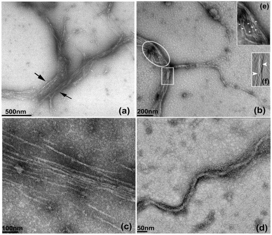
Figure 5. TEM images of MamK filaments.
MamK was negatively stained with 2% uranyl acetate and incubated with 0.2 mM ATγP. Incubation of filaments with the nucleotide analogue generated filaments of various sizes (Figure 5a–d). A parallel arrangement of filaments of (∼11–18 nm) was observed after 15 min of incubation (Figure 5c); filament bundling of the order ∼80–270 nm s (Figure 5b). Image 5b also showed the alignment of numerous filaments (circled) in the process of forming bundles composed of smaller filaments of ∼4–12 nm (enlarged; Figure 5e inset) and twisted rope-like morphologies of width ∼38 nm (square) composed of smaller filaments of ∼12–16 nm; (enlarged; Figure 5f inset); a network of individual filaments (16–18) nm in the process of forming bundles of 50–100 nm (Figure 5a) and longer filaments (25–30 nm) of length >2.0 µm (Figure 5d). Figures 5a, 5b and 5d were imaged after 30 mins.

