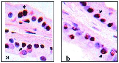
Figure 1.
Immunohistochemical demonstration of ERβ in ventral prostates of rats and mice. Frozen sections of ventral prostates from mice (a) and rats (b) stained for ERβ with anti-ERβ, 503-IgY. Positive immunoreaction is indicated by a brown color, and typical examples are marked by arrows. Stromal cell nuclei and some epithelial cell nuclei that are negative for ERβ are made evident by the blue counterstain.

