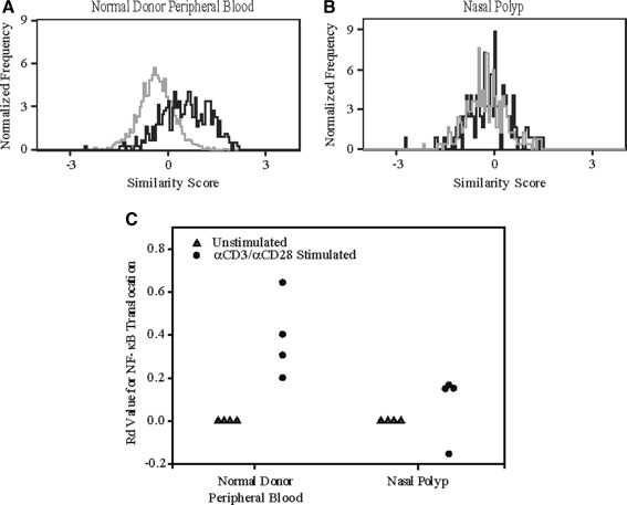
FIG. 3.
Nasal polyp-derived T cells show impaired ability to translocate NF-κB as determined by multispectral imaging flow cytometry. Following stimulation for 1 h with anti-CD3/CD28, T cells derived from the nasal polyp were deficient in their ability to translocate NF-κB compared to T cells from normal donor PBL. A In normal donor PBL, the stimulated similarity distribution plot (black) for NF-κB is shifted to the right of the unstimulated plot (gray), demonstrating translocation. B The similarity distribution plots for the unstimulated and stimulated samples in the nasal polyp completely overlay, indicating the cells were unable to translocate NF-κB with stimulation. C The Rd values after activation for the T cells derived from normal donor PBL were significantly greater than for T cells derived from nasal polyps (n = 4, p < 0.05).

