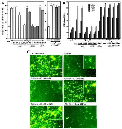
Figure 4.
Effect of HN on Aβ-induced cell death in primary neurons. (A and B) Primary cortical neurons were treated with 25 μM Aβ1–43 in the presence or absence of sHN or its derivatives. In A, cell mortality was measured 72 h after Aβ treatment with or without HN peptides. Neurons were similarly treated, as a positive control, with 20 μM etoposide in the presence or absence of 10 μM HN peptides for 72 h. In B, the LDH activity in the culture media was monitored by sampling 6 μl of the media culturing neurons treated with 25 μM Aβ1–43 in the presence or absence of HN peptides at 24, 48, or 72 h after the onset of Aβ treatment. The LDH release was also measured in neurons treated with 20 μM etoposide in the presence or absence of HN peptides at 24, 48, or 72 h after the onset of etoposide treatment. These experiments were performed independently for both assays. (C) Fluorescence microscopic views by Calcein-AM staining for neuronal viability. Seventy-two hours after treatment with 25 μM Aβ1–43, in the presence or absence of HN peptides, neurons were stained with Calcein-AM. Insets indicate magnified views of similarly treated independent cultures. Representative views are indicated.

