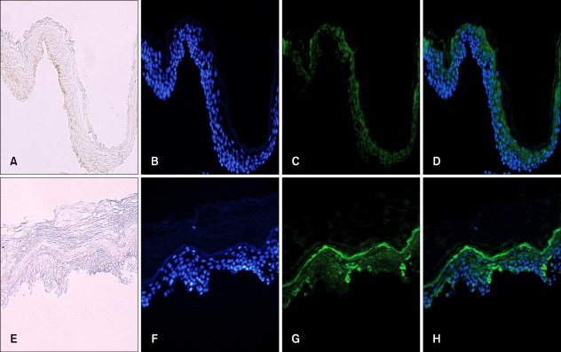
Fig. 3.
Immunofluorescence staining of paired suction-blistered epidermis samples with anti-tumor necrosis factor (TNF)-α antibody. (A, E) without staining; (B, F) staining of nuclei with Hoechst 33258; (C, G) staining with anti-TNF-α antibody; (D, H) staining with anti-TNF-α antibody (green) and counterstaining of nuclei with Hoechst 33258 (blue) (A~D) in the normally pigmented epidermal specimens. Cells were weakly stained with anti-TNF-α antibody. (E~H) in the depigmented epidermal specimens. More cells stained strongly with anti-TNF-α antibody. The strong TNF-α-positive cells were mainly found in the lower layer (A~H, ×200).

