Figure 1.

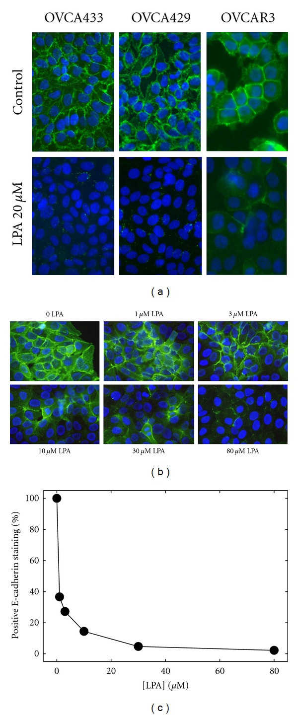
LPA induces E-cadherin junction disruption in EOC cells. (a) Confluent monolayers of OVCA433, OVCA429, or OVCAR3 cells, as indicated, were treated with LPA (20 μM) for 18–24 hours and processed for immunofluorescence staining for E-cadherin using anti-E-cadherin ectodomain antibody (1 : 300) and Alexa Fluor 488-conjugated secondary antibody (1 : 500; green). Blue-DAPI-stained nuclei. (b) To evaluate the dose-dependence of LPA-induced junction loss, OVCA429 cells were treated with LPA at the concentrations indicated for 24 hours and processed for E-cadherin immunofluorescence (green) as described in (a) above. Blue-DAPI-stained nuclei. (c) Junction loss was quantified by counting the number of cells/field with two remaining E-cadherin immunostained borders in a minimum of 5 fields per treatment (at least 100 cells).
