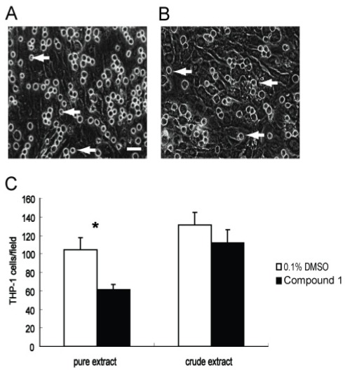
Figure 4.
Blocking adhesion of THP-1 cells onto a monolayer of WI26 cells by compound 1. Representative pictures of the adhering cells taken with 400 fold magnification. Equal numbers of THP-1 cells with 1 (30 μg/mL) or 0.1% DMSO were added to a monolayer of WI26 cells in triplicate wells of 24-well plates, respectively. Adhering THP-1 cells after 3-h-incubation were counted in four random visual fields of each well. (A) The attachment of THP-1 cells (marked by arrows) onto confluent WI26 cells, treated with 0.1% DMSO; (B) The attachment of THP-1 cells onto confluent WI26 cells, treated with 1 (30 μg/mL). It is clear from these images that adhesion of THP-1 cells was significantly blocked by 1. Bar = 25 μm. Each point represents the mean ± SD of three independent experiments. Significant difference from the value of 0.1% DMSO solvent control was marked, *p < 0.05.

