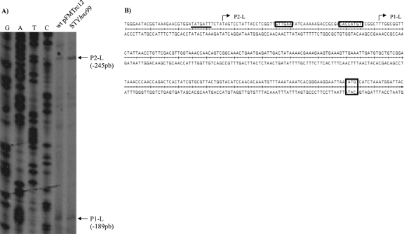
Fig 7.
dsbL promoter region in N-minimal medium. (A) Primer extension assay results for dsbL, performed in N-minimal medium at 12 h from S. Typhi IMSS-1 wild type with pFMTrc12 and from IMSS-1 STYhns99, both containing the assT+78/dsbL+105 gene fusion. (B) Upstream regulatory sequence of dsbL. The locations of the two transcription start sites (P1-L and P2-L) are shown. The putative −10 and −35 σ32 promoter sequences for P1-L are shown in rectangles, whereas the −10 TATA box for the P2-L σ70 promoter is underlined. The initiation codon for the dsbL gene is indicated in a rectangle.

