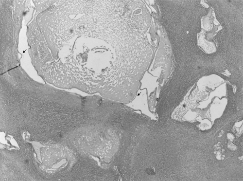
Fig 2.
Histopathology of liver lesion. One central cyst and budding daughter cysts with a trilayered membrane wall (arrows) are visible. No protoscolices, hooklets, or calcareous corpuscles are visible; however, the image is still compatible with echinococcosis. Magnification, ×100; hematoxylin and eosin stain.

