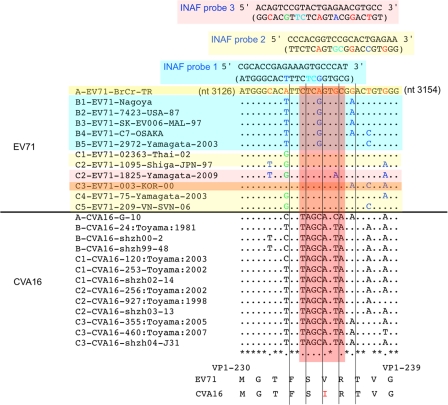
Fig 3.
Comparison of the nucleotide sequences of the EV71 and CVA16 genomes for the INAF probes. Genomic sequences of EV71 and CVA16 strains are shown with their genotypes and names. Nucleotide differences in the EV71 genomic sequences and in INAF probes are highlighted in red (genotype A), blue (genotype B), and green (genotype C). Flanking nucleotides of oxazole yellow dye are colored in cyan in the INAF probe sequences. INAF probes (1, 2, and 3) and the target EV71 strains are colored in cyan, yellow, and pink, respectively. For detection of genotype C3 of EV71, INAF probes 2 and 3 were required. The amino acid sequences of EV71 and CVA16 in the INAF probe-binding region are shown below.

