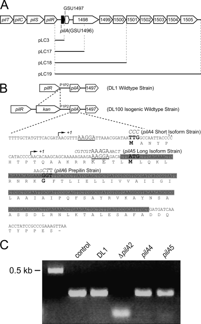
Fig 1.
(A) Genomic organization of the pilus biosynthesis genes and gene cluster downstream of pilA (GSU1496). The black bars indicate DNA cloned into the plasmids constructed for complementation experiments. (B) Sequences of the pilA gene and PilA protein, including the mutations introduced for each strain used in this work. In the isogenic wild-type and all mutant strains, a kanamycin cassette was inserted upstream of the P1 and P2 promoter regions. The arrows indicate the two transcription initiation sites. The two ribosome-binding sites are underlined. The translation start codons (TTG and ATG) and the peptidase cleavage site (GGT) are in boldface. The base changes leading to amino acid replacements are depicted in italics for each mutant strain. The deleted sequence of the pilA gene in the ΔpilA2 strain is highlighted in gray. (C) DNA gel of semiquantitative reverse transcriptase PCRs. (1) proC control; (2) wild-type pilA in DL1; (3) ΔpilA2; (4) short-isoform (pilA4) strain; and (5) long-isoform (pilA5) strain.

