Fig 3.
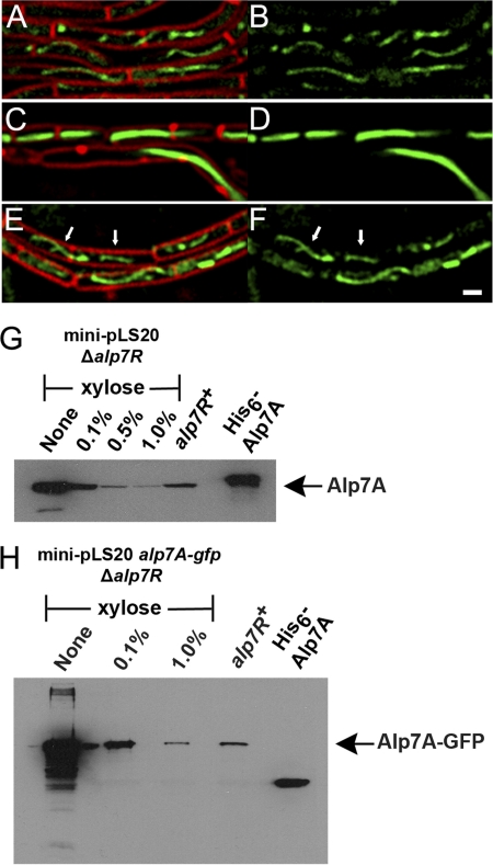
Depletion of Alp7R leads to overproduction of Alp7A. (A to F) Fluorescence microscopy images of strains grown in the presence or absence of d-xylose (agarose pads; scale bar equals 1 μm; all panels are at the same scale). (A and B) JP3196 (PY79 carrying mini-pLS20 alp7A-gfp) in the absence of xylose. (C to F) JP3245 (PY79 with an integrated xylose-inducible copy of alp7R, carrying mini-pLS20 alp7A-gfp Δalp7R) in the absence of xylose (C and D) or in the presence of 0.5% xylose (E and F). Arrows denote filaments whose dynamic behavior can be tracked in the lower left section of the corresponding movie (see Fig. S2 in the supplemental material). (A, C, and E) Cell membranes (FM4-64) and GFP. (B, D, and F) GFP only. See also Fig. S1, S2, and S3 in the supplemental material. (G and H) Immunoblots prepared from lysates of JP3302 (PY79 with an integrated xylose-inducible copy of alp7R, carrying mini-pLS20 Δalp7R) (G) or JP3245 (PY79 with an integrated xylose-inducible copy of alp7R, carrying mini-pLS20 alp7A-gfp Δalp7R) (H) grown in the presence of various concentrations of xylose. The filter was probed with a polyclonal anti-Alp7A serum. Lanes are labeled with the xylose concentrations used. The lane labeled “alp7R+” shows the steady-state level of Alp7A that is present in JP3133 (PY79 carrying mini-pLS20) (G) or of Alp7A-GFP that is present in JP3196 (PY79 carrying mini-pLS20 alp7A-gfp) (H). The rightmost lane contains purified His6-Alp7A (G and H).

