Fig 4.
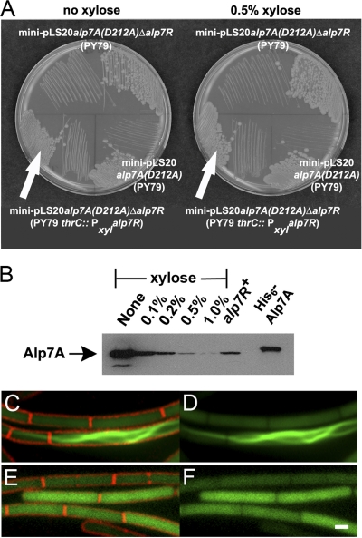
Deletion of alp7R causes Alp7A(D212A) to assemble into filaments. (A) LB agar plates containing 10 μg ml−1 tetracycline with or without 0.5% d-xylose were incubated overnight at 30°C. Lower left, JP3247 [PY79 with an integrated xylose-inducible copy of alp7R, carrying mini-pLS20 alp7A(D212A) Δalp7R]; lower right, JP3169 [PY79 carrying mini-pLS20 alp7A(D212A)]; top, JP3248 [PY79 carrying mini-pLS20 alp7A(D212A) Δalp7R]. (B) Immunoblot prepared from lysates of JP3247 grown in the presence of various concentrations of xylose. The filter was probed with a polyclonal anti-Alp7A serum. Lanes are labeled with the xylose concentrations used. The lane labeled “alp7R+” shows the steady-state level of Alp7A(D212A) that is present in JP3169. The rightmost lane contains purified His6-Alp7A. (C to F) Fluorescence microscopy images of strain overproducing Alp7A(D212A) (agarose pads; scale bar equals 1 μm, all panels are at the same scale). JP3233 [PY79 with an integrated xylose-inducible copy of alp7R, carrying mini-pLS20 alp7A(D212A)-gfp Δalp7R] in the absence of xylose (C and D) or in the presence of 0.5% xylose (E and F). (C and E) Cell membranes (FM4-64) and GFP. (D and F) GFP only. See also Fig. S4 in the supplemental material.

