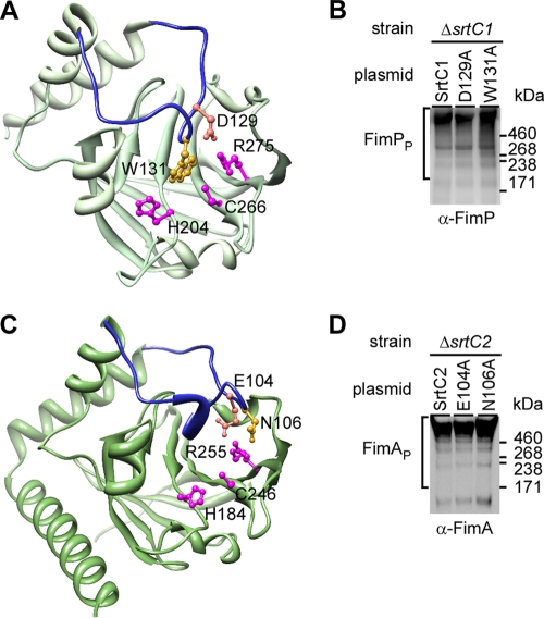
Fig 5.
Analysis of a flexible lid in Actinomyces sp. fimbria-specific sortases. (A) A lid (blue) with anchor residues D129 and W131 covering catalytic residues H204, C266, and R275 is shown in the three-dimensional (3D) crystal structure of Actinomyces sp. fimbria-specific sortase SrtC1. Shown in panel C is a 3D structure of Actinomyces fimbria-specific sortase SrtC2, as modeled after the SrtC2 structure. (B and D) Western blotting for fimbrial polymerization of Actinomyces sp. cells expressing wild-type SrtC1 and its lid mutants (B) or wild-type SrtC2 and its lid mutants (D) was carried out as described in the legend to Fig. 1.

