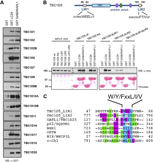
Fig 1.
TBC proteins bind human ATG8 proteins via canonical LIR. (A) ATG8-interacting TBC proteins. HEK 293T cell lysates overexpressing GFP-tagged human TBC proteins were subjected to pulldown assays using GST, GST-MAP1LC3A, and GST-GABARAPL1. Coprecipitated TBC proteins were detected with anti-GFP antibody. Inputs are shown in Fig. S1A in the supplemental material. WB, Western blot. (B) (Top) Schematic representation of TBC1D5 with Pfam domains annotated and LIR motifs identified. (Bottom) The wild type and catalytic RA and LIR mutants of TBC1D5 were transiently expressed in 293T cells and subjected to pulldown assays with GST alone or GST-ATG8 proteins. GST fusion proteins were visualized by Ponceau S staining. (C) Alignments of canonical LIR motifs from annotated ATG8-interacting partners and LIR motifs mapped in TBC1D5. Magenta, acidic residues; blue, small and hydrophobic residues; green, serine residues.

