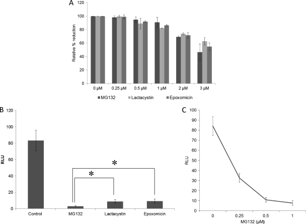
Fig 1.
The proteasome inhibitors significantly reduced the levels of HEV replication. (A) Toxicity of inhibitors to Huh7 S10 cells. The results shown are from an alamarBlue assay for Huh7 S10 cells treated with the inhibitors MG132, lactacystin, and epoxomicin. The assay was performed on the fourth day after treatment. The concentrations of inhibitors are indicated. Mean values from three independent experiments are plotted. (B) HEV replication is reduced by treatment with proteasome inhibitors. Relative luciferase activities are shown for Huh7 S10 cells transfected with capped RNA transcript of the pSK-HEV2RLuc clone. Inhibitor treatment started 1 day posttransfection, and the concentration of inhibitors was 1 μM. A luciferase assay was performed on the fifth day posttransfection. Mean values from six independent experiments are plotted. Statistical analysis was performed using analysis of variance followed by Dunnett's procedure, and significance was set at a P level of <0.05 (indicated with an asterisk). Data analysis was performed using JMP9 software. (C) Effect of MG132 on HEV replication. Relative luciferase activities are shown for Huh7 S10 cells transfected with capped RNA transcript of the pSK-HEV2RLuc clone. Inhibitor treatment started 1 day posttransfection, and the concentration of MG132 used is indicated. A luciferase assay was performed on the fifth day posttransfection. Mean values from three independent experiments are plotted.

