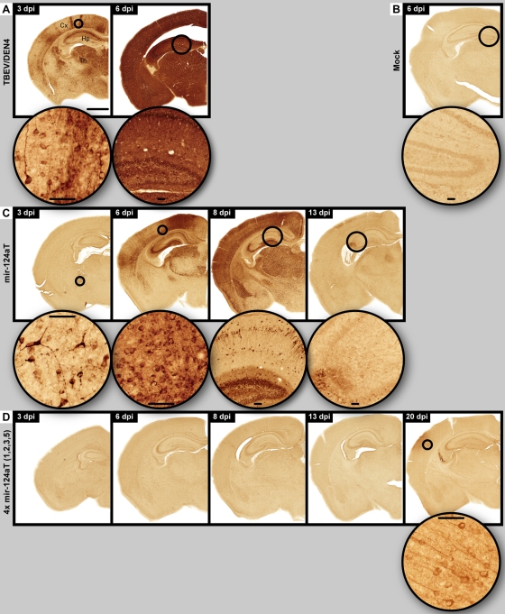
Fig 5.
Spatiotemporal distribution of viral antigens in neurons. Immunohistochemistry for viral antigens was performed on CNS of mice infected with the parental TBEV/DEN4 (A), mir-124aT (C), or 4x mir-124aT(1,2,3,5) (D) virus on the indicated day p.i. (dpi). A mock control is shown in panel B. The bar (1,000 μm) (in panel A) applies to all panels showing a whole-mouse brain hemisphere. Round insets show circled areas of cortex (Cx), thalamus (Th), or hippocampus (Hp) at higher magnification (bars in insets, 50 μm).

