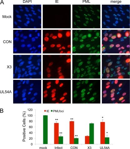
Fig 8.
Cells pretreated with X3 siRNA retain PML in cellular PML bodies following infection. HEL fibroblast cells were untransfected or transfected with X3, UL54A, or CON siRNAs 24 h prior to infection with HCMV at an MOI of 1.0. Cells were fixed at 48 hpi, and PML was detected by immunostaining. (A) Images show the localization of IE and PML proteins. Cells with typical PML bodies are seen in normal cells (Mock). PML protein was observed as diffuse nuclear staining in HCMV-infected cells pretreated with siCON. DAPI staining is used to define nuclei. (B) The percentages of fibroblasts with IE-positive staining or PML bodies were plotted. Over 200 cells were scored per sample. Histograms show the averages of three independent experiments, and the error bars denote the standard deviations. *, P < 0.0004 relative to X3; **, P < 0.0001 relative to X3.

