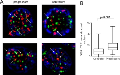
Fig 2.
Colocalization of the DNA damage response factor 53BP1 and TRF1 in HIV-1-specific CD8 T cells. Lymphocytes were labeled with anti-53BP1 (red) and anti-TRF1 (green) antibodies; DAPI was used as a nuclear counterstain. (A) Representative examples of 53BP1 and TRF1 staining in sorted tetramer-positive HIV-1-specific CD8 T cells. The arrows indicate costaining of TRF1 and 53BP1 as TIF. The images are shown at a magnification of ×100. (B) Cumulative analysis of 53BP1/TRF1 colocalization in HIV-1-specific CD8 T cells. A total of 120 cell nuclei (a single focal plane for each nucleus) from progressors and controllers (n = 5 each) were analyzed. Significance was tested using a two-sided, unpaired t test.

