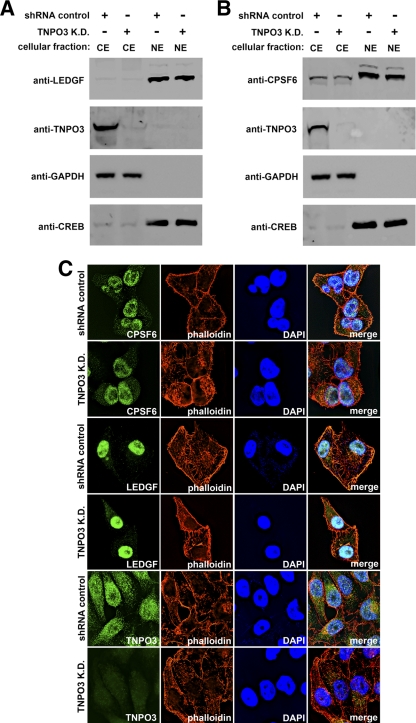
Fig 3.
Subcellular localization of LEDGF and CPSF6 in TNPO3-depleted cells. (A and B) To determine the cellular localization of LEDGF and CPSF6, HeLa TNPO3 knockdown cells (TNPO3 K.D.) or HeLa cells transduced with the empty vector pLKO.1 (shRNA control) were biochemically fractionated to separate nuclear from cytosolic proteins. Cytosolic extracts (CE) and nuclear extracts (NE) were blotted using antibodies against LEDGF, CPSF6, and TNPO3. To assay the bona fide origin of the extracts, the fractions were Western blotted using anti-glyceraldehyde-3-phosphate dehydrogenase (anti-GAPDH) and anti-CREB as cellular markers for the cytosol and nucleus, respectively. (C) Subcellular localization of CPSF6 and LEDGF by immunofluorescence. HeLa TNPO3 knockdown cells (TNPO3 K.D.) or HeLa cells transduced with the empty vector pLKO.1 (shRNA control) were fixed and stained using antibodies against CPSF6, LEDGF, and TNPO3. The nuclear compartment and F-actin was labeled using 4′,6-diamidino-2-phenylindole (DAPI) and phalloidin, respectively. Similar results were obtained in three independent experiments, and a representative experiment is shown.

