Fig 2.
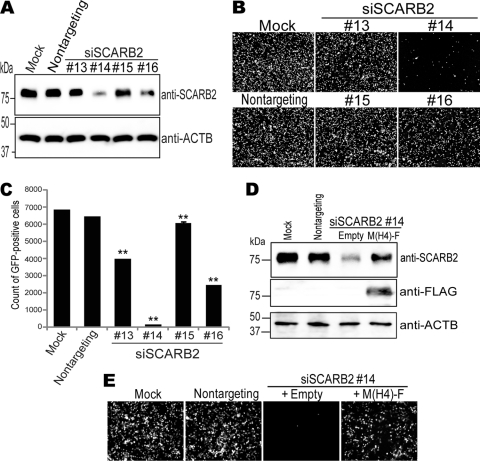
Downregulation of SCARB2 expression inhibited EV71-GFP infection. (A) Cell lysates were prepared from RD cells mock treated or treated with nontargeting siRNA or siSCARB2 #13, #14, #15, or #16 and were analyzed by Western blotting using an anti-SCARB2 antibody or an anti-ACTB (β-actin) antibody. (B and C) The siRNA-treated RD cells were infected with EV71-GFP and imaged via fluorescence microscopy at 24 h postinfection (B) and then analyzed by FACS to quantify the number of GFP-positive cells (C). A total of 10,000 cells were analyzed by FACS, and data are shown as mean counts with SD (n = 3). (D) SCARB2 expression was restored by exogenous M(H4)-F expression. Cell lysates were prepared from RD cells mock treated or treated with nontargeting siRNA or siSCARB2 #14, which were transfected with either empty vector or pCA-M(H4)-F and analyzed by Western blotting using an anti-SCARB2 antibody, an anti-FLAG antibody, or an anti-ACTB antibody. (E) M(H4)-F expression rescued the EV71-GFP infection that was inhibited by treatment with siSCARB2 #14. The siRNA-treated RD cells with/without plasmid transfection were infected with EV71-GFP. After 24 h, the cells were imaged via fluorescence microscopy.

