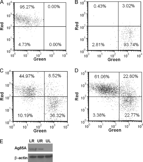
Fig 5.
Apoptotic bodies from MVA-infected cells are phagocytized by noninfected DC. PKH-67-labeled ALDC were infected with rMVA-Ag85A (MOI = 3) as described in Materials and Methods and cultured overnight at 37°C. PKH-46-labeled autologous ALDC were then added to the culture, incubated at either 4°C or 37°C for 4 h, and analyzed by flow cytometry. (A) PKH-46-labeled (red) cells. (B) PKH-67-labeled (green) cells. (C) Mixed culture incubated at 4°C. (D) Mixed culture incubated at 37°C. Dot plots shown are gated on FSChigh MHCII+ CD11c+ DEC205+ live single events and are representative of experiments performed using cells from 10 individual animals. (E) Western blot (representative of 3 independent experiments) showing the presence of Ag85A and β-actin in FACS-sorted ALDC from panel D. LR, lower right; UR, upper right; and UL, upper left.

