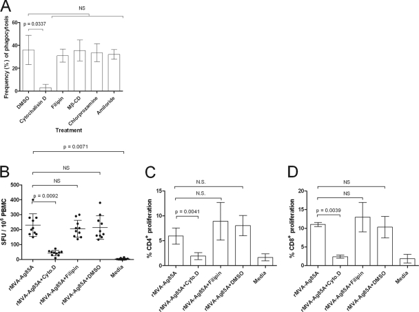
Fig 7.
Uptake of apoptotic bodies by ALDC is by actin-mediated phagocytosis. (A) PKH-67-labeled ALDC were infected with rMVA-Ag85A (MOI = 3) as described in Materials and Methods. PKH-46-labeled autologous ALDC were added to the culture, incubated at 37°C for 4 h in the presence of the indicated inhibitors, and analyzed by flow cytometry. Cells were gated on FSChigh MHCII+ CD11c+ DEC-205+ live single events. Bars represent means (n = 10) of results from events present in the upper right (UR) quadrant as shown in Fig. 5 (double positive for PKH-67 and PKH-46). Error bars indicate standard deviations. (B, C, and D) ALDC were infected with rMVA-Ag85A (MOI = 3), and after an overnight culture, autologous ALDC were added. Antigen presentation was measured by ELISPOT assay (B) or T cell proliferation by flow cytometry (C and D). Bars represent means of results from 10 animals analyzed in duplicate, and error bars indicate standard deviations. NS, not significant.

