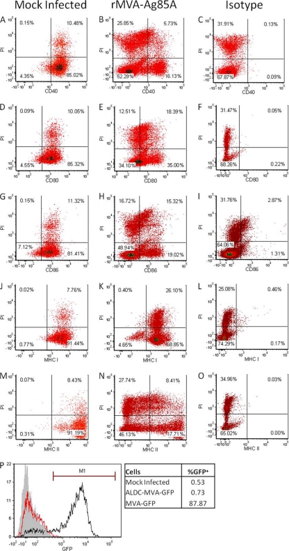
Fig 8.
Infection by MVA downregulates expression of costimulatory molecules, MHC-I, and MHC-II in both infected and noninfected ALDC. ALDC (1 × 10⁶) were mock infected (A, D, G, J, and M) or infected with rMVA-Ag85A (B, C, E, F, H, I, K, L, N, and O) using a suboptimal MOI of 0.5 PFU/cell at 4°C for 60 min to allow virus attachment; excess virus was removed by extensive washing, and the same number of uninfected autologous ALDC were added. After a 16-h culture at 37°C, the cells were harvested and stained with propidium iodide and mouse anti-bovine MAbs or isotype control antibodies and cell surface expression was analyzed by flow cytometry. Events shown were gated on single FSChigh DEC-205+ events. Dot plots are representative of 3 independent experiments. (P) ALDC were mock infected (gray histogram) or infected (red histogram) with rMVA-GFP and washed as described above. Cells were then cocultured with chicken embryo fibroblasts (CEF) for 16 h at 37°C. Cells were then harvested, and GFP expression was measured by flow cytometry. The black histograms represent CEF infected with rMVA-GFP (MOI = 1). The overlay is representative of 3 independent experiments.

