Fig 1.
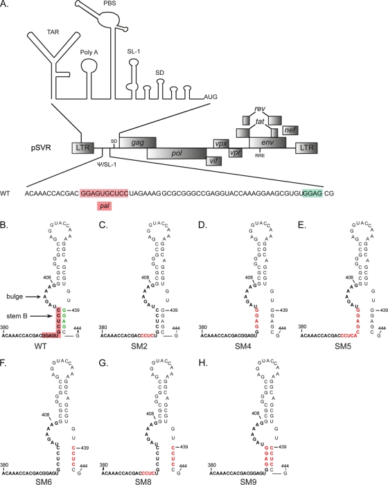
Structure of stem-loop 1 in wild-type and mutant HIV-2. (A) Schematic of the molecular clone pSVR of the HIV-2 ROD genome, with the structure of the 5′ leader RNA (20) shown above and the sequence of the packaging signal (Psi)/stem-loop 1 (SL-1) region detailed below. The palindrome pal (residues 392 to 401) and the GGAG motif at position 439 are highlighted in red and green, respectively. TAR, trans-activator response; PBS, primer binding site; SD, major splice donor. (B to H) Structures of SL-1 in the WT (B) and substitution mutants (C to H) based on the previously described structure of SL-1 (4). The palindrome pal is highlighted in red on the WT structure. Stem B and the distal bulge (residues 402 to 409) are indicated. SM2 (C) (residues 392 to 395) has been described and characterized previously (41). Red letters represent mutated residues.

