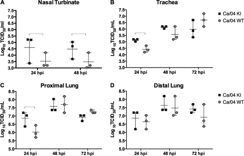
Fig 2.
Replication of PB1-F2 recombinant viruses in porcine respiratory explants. Explants from nasal turbinates (A), trachea (B), and proximal (C) and distal (D) lung were infected with 106 TCID50 of either Ca/04 WT or KI. The bathing medium was collected at the indicated time points and titrated by the TCID50 method in MDCK cells. Values shown are the mean and range of virus titers (log10 TCID50/ml) obtained from triplicate explant preparations. A two-tailed Student t test was used to determine significant differences between two treatment means for each data point. Dashed brackets indicate statistically significant differences (P < 0.05).

