Fig 3.
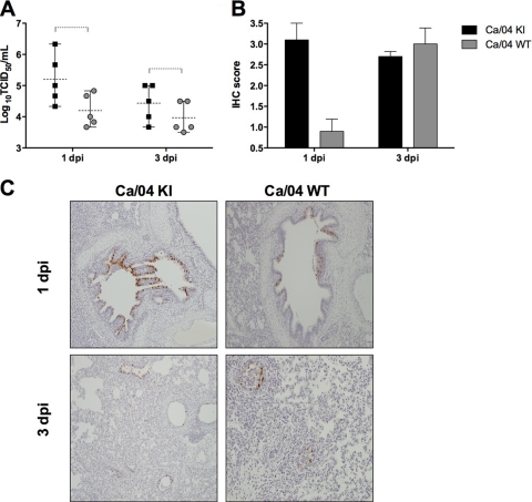
Replication and immunohistochemical analysis of PB1-F2 recombinant viruses in swine lungs. Groups of pigs (n = 10) were infected with 105 TCID50 of PB1-F2 recombinant viruses, and five animals from each group were euthanized either at 1 or 3 dpi. The lungs were collected and processed for virus titration and immunohistochemical analysis. (A) Pulmonary replication of PB1-F2 isogenic viruses in pigs. Values are means and ranges of virus titers (log10 TCID50/ml) in bronchoalveolar lavage fluid (BALF) collected at the indicated time points. Two-way ANOVA was used to determine significant differences between two treatment groups. Dashed brackets indicate statistically significant differences (P < 0.05). (B) Immunohistochemical staining against influenza A virus nucleoprotein (NP) in the lungs of infected pigs. Values given are the mean ± the standard error of the mean IHC scores based on the percentage of influenza virus-positive cells in the airway and lung interstitium. (C) Representative IHC slides depicting viral antigen primarily in airway epithelium at 1 and 3 dpi. Scattered labeling in the interstitium at 3 dpi is present.

