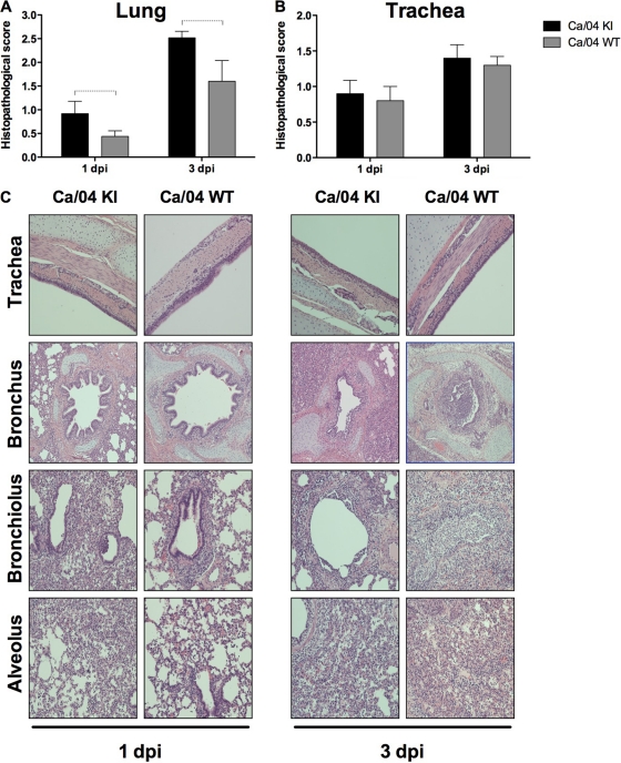
Fig 5.
PB1-F2 exacerbates microscopic pneumonia in swine. Groups of pigs (n = 10) were infected with 105 TCID50 of PB1-F2 recombinant viruses. At 1 and 3 dpi, five animals from each group were euthanized, and the histopathological changes in the lower respiratory tract were evaluated. (A) Histopathologic scores in the lungs. The differences are statistically significant (two-way ANOVA, P < 0.05). (B) Histopathologic scores of trachea. The differences are not statistically significant (two-way ANOVA, P > 0.05). (C) Photomicrographs representing microscopic pneumonia in Ca/04 KI and Ca/04 WT virus. Dashed brackets indicate statistically significant differences (P < 0.05).

