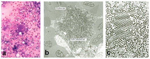
Figure 1.
(a) Giemsa-stained impression smears of the spleen of RSIV-infected red sea bream display enlarged cells characterized by basophilic staining. (b) Electron micrograph of RSIV‑infected spleen cells. (c) Higher magnification of the virions seen in panel B. All photographs were kindly provided by Dr. K. Inouye.

