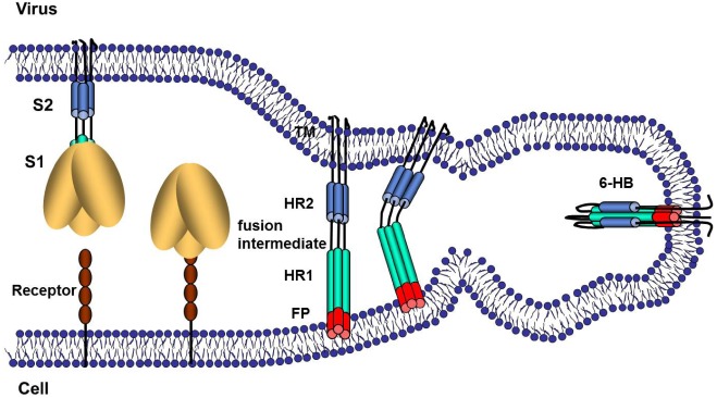
Figure 2.
Schematic illustration of CoV S protein-mediated membrane fusion. The illustrations represent several steps of S protein conformational changes that may take place during membrane fusion. In the first step, receptor binding, pH reduction and/or S protein proteolysis induces dissociation of S1 from S2. This step is documented for some MHVs [29,30]. In the second step, the fusion peptide (FP) is intercalated into the host cell membrane. This is the fusion-intermediate stage. In the third stage, the part of the S protein nearest to the virus membrane refolds onto a heptad repeat 1 (HR1) core to form the six-helix bundle (6-HB), which is the final postfusion configuration of the S2 protein.

