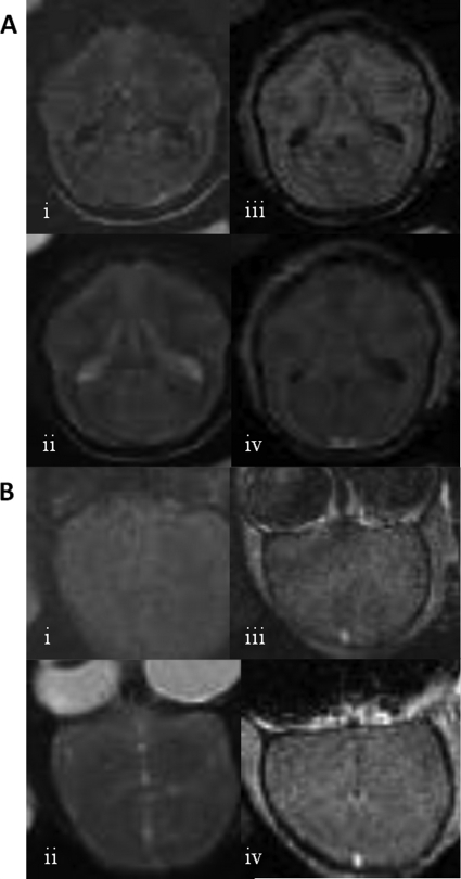
Fig 5.
Shown are MRI images of two different A. nancymae animals after inoculation with M002, (A) Animal 1. One month prior, 1.2 × 10⁸ PFU of M002 was inoculated in the right frontal lobe. Images are in the axial plane. Shown are (i) FLAIR, (ii) T2-weighted, (iii) T1-weighted pregadolinium, and (iv) T1 postgadolinium images. (B) Animal 2. Seven months prior, 4.8 × 10⁸ PFU of M002 was inoculated in the right frontal lobe. Images are in coronal plane and inverted. Shown are (i) FLAIR, (ii) T2-weighted, (iii) T1-weighted postgadolinium, and (iv) additional plane, T1 postgadolinium images. No pathological changes are seen after M002 administration.

