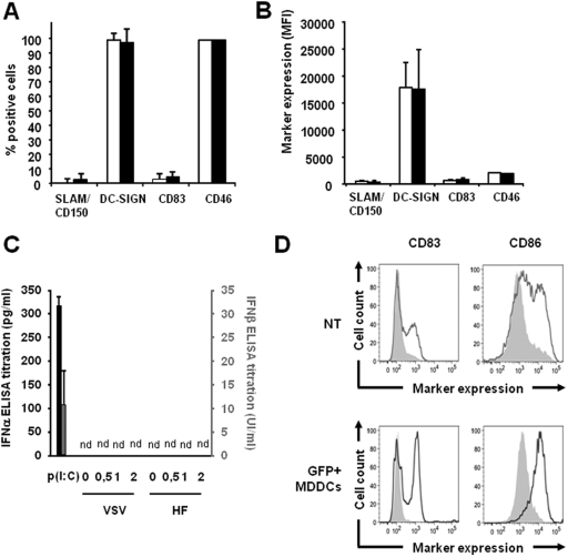
Fig 5.
No difference in the expression of MV receptors and activation or maturation markers of MDDCs occurs upon transduction with H/F-LVs. (A or C) Results for MDDC phenotypes upon H/F-LV transduction. (D) Phenotypic analysis of poly(I:C)-matured MDDCs with or without transduction. In panels A and B, the percentages (A) and MFI values (B) for positive immature MDDCs for the CD150/SLAM, DC-SIGN, CD83, and CD46 molecules were evaluated by flow cytometry before (white bars) and after (black bars) transduction with a GFP-encoding H/F-LV (MOI of 10 for both). These analyses were performed using the MDDC subpopulation. The results are presented as means ± the SEM (n = 4). (C) IFN-α (black bars) and IFN-β (gray bars) were quantified by ELISA in the supernatants of MDDCs upon transduction with H/F-LVs or VSV-G-LVs at the indicated MOIs. Supernatants were harvested at 24 h posttransduction. Treatment with poly(I:C) at 50 μg/ml was used as a positive control for type I IFN secretion by MDDCs. nd, not detected (neither IFN-α nor IFN-β was recovered in the culture supernatants). The results are presented as means ± the SEM (n = 2). (D) Comparison of CD83 and CD86 expression levels on untransduced (NT) versus H/F-LV-transduced MDDCs. The cells were transduced with a GFP-encoding LV (MOI = 10). GFP-positive MDDCs were positively selected by FACS to achieve nearly 100% transduced cells. Untransduced and GFP+ cells were subcultured with (thick line) or without (gray filled) poly(I:C) (a TLR3 agonist) at 12.5 μg/ml and subsequently stained for the CD83 and CD86 maturation markers (n = 3).

