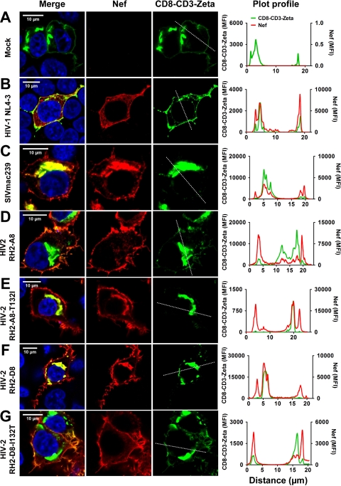
Fig 9.
Subcellular distribution of CD8-CD3ζ in the presence of wild-type and mutant HIV-2 Nef proteins. HEK293T cells cotransfected with equal amounts of plasmid DNA encoding a CD8-CD3ζ-GFP fusion and the indicated nef alleles were analyzed by confocal microscopy. CD8-CD3ζ was detected by GFP, whereas HA-tagged Nef proteins were detected by indirect immunofluorescence (anti-HA mouse monoclonal antibody followed by secondary Alexa Flour 568-conjugated goat anti-mouse IgG antibody). Merged and single images for various Nef alleles and CD8-CD3ζ-GFP are shown for mock (A), (B) HIV-1 NL4-3 Nef, (C) SIVmac239 Nef, (D) HIV-2 RH2_A8 Nef, (E) HIV-2 RH2_A8-T132I Nef, (F) HIV-2 RH2_D8 Nef, and (G) HIV-2 RH2_D8-I132T samples. The last panel under each condition shows the mean fluorescence intensity at the ROI drawn across the cells.

