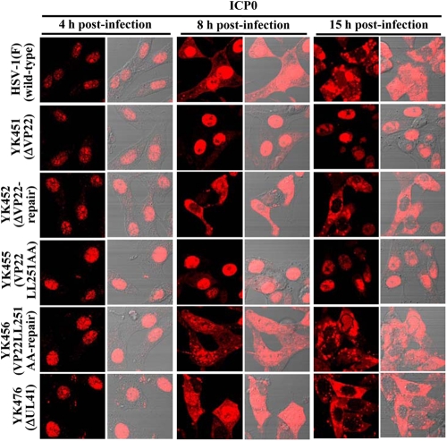
Fig 12.
Digital confocal microscope images showing the localization of ICP0 in Vero cells infected with wild-type HSV-1(F), YK451 (ΔVP22), YK452 (ΔVP22-repair), YK455 (VP22LL251AA), YK456 (VP22LL251AA-repair), or YK476 (ΔUL41) at an MOI of 1. Infected Vero cells were fixed at the indicated times postinfection, permeabilized, stained with an antibody to ICP0, and examined by confocal microscopy. Left and right columns at each time point show protein fluorescence and simultaneous acquisition of protein fluorescence and digital interference contrast, respectively.

