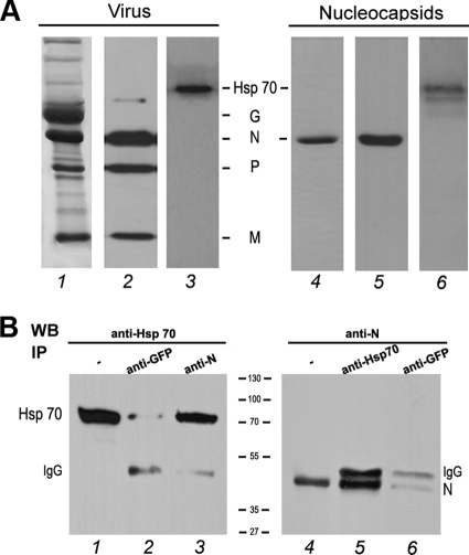
Fig 1.
Hsp70 interacts with the rabies virus N protein. (A) Hsp70 is detected in purified virus and in purified nucleocapsid. Purified virus (5 μg) and purified nucleocapsid (5 μg), prepared as described in Materials and Methods, were analyzed by SDS-PAGE followed by Coomassie blue staining (lanes 1 and 4) and Western blotting using a mixture of anti-N, anti-P, and anti-M antibodies (lanes 2 and 5) and a polyclonal anti-Hsp70 (lanes 3 and 6). (B) BSR cells were cotransfected with plasmids expressing Hsp70 and rabies N protein. Two days after transfection, cells were harvested and lysed. Cell lysate was incubated with the irrelevant polyclonal anti-GFP antibody (lanes 2 and 6), with the mouse MAb anti-N antibody (lane 3), or with the polyclonal anti-Hsp70 antibody (lane 5). Immune complexes were analyzed by Western immunoblotting using anti-Hsp 70 (lanes 2 and 3) and anti-N (lanes 5 and 6) antibodies. Direct cellular extracts of transfected cells (input) also were analyzed by WB with anti-Hsp70 (lane 1) and anti-N (lane 4). Bands corresponding to Hsp70, N, and heavy-chain IgG are indicated.

