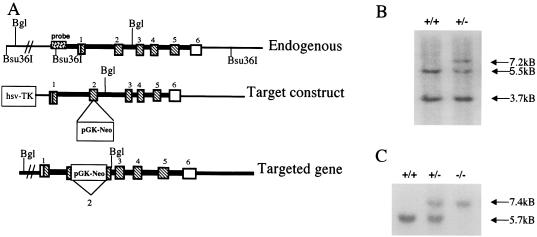
Figure 1.
SP-C targeting construct and Southern blot identification of targeted ES cells and mice. (A) A restriction map of the SP-C gene is shown above the targeting vector. Exons are numbered 1–6. The bottom line diagrams the targeted SP-C gene. Precise homologous recombination between lines 1 and 2 results in exon 2 interrupted by the neomycin resistance (pGKneo) gene and loss of the nonhomologous herpes simplex virus thymidine kinase (HSV-TK) gene (bottom line). (B) Southern blot analysis of Bsu36I genomic DNA from ES cells. Bsu36I-digested DNA was probed with a 5′ Sph-Pst fragment adjacent to the targeting DNA, which detects 3.7-kb upstream and 5.5-kb downstream endogenous bands (+/+, lane 1). DNA from a targeted ES cell colony (+/−) shows the 5.5-kb allele and the targeted allele that are increased by the size of the inserted pGKneo cassette to 7.2 kb. (C) Southern blot analysis of genomic DNA from SP-C gene-targeted mice. The 5′ probe was used for blots of BglII-digested DNA. A single, 5.7-kb BglII band is detected in wild-type mice by the 5′ probe. Two bands are detected in mice heterozygous for the targeted site (+/−). Homozygous SP-C (−/−) mice produce only the 7.4-kb, larger upper band, indicating that both SP-C alleles are interrupted.

