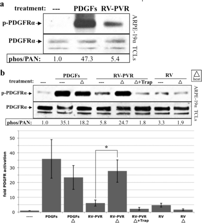
Fig 1.
Vitreal PDGFs were inhibited by a heat-labile agent. (a) PVR vitreous activated PDGFRα poorly. ARPE-19α cells, which stably express PDGFRα and induce experimental PVR (42), were grown to near confluence and serum starved overnight. The cells were then treated with serum-free medium alone (—), PDGFs totaling 75 ng/ml (comprising the A, AB, and B isoforms at 40, 30, and 5 ng/ml, respectively, which reflects the composition of PDGFs in PVR vitreous) (39), or vitreous (0.2 ml) from rabbits with PVR (RV-PVR). After 5 min of incubation at 37°C, cells were lysed, and the resulting total cell lysates (TCLs) were subjected to anti-phospho-PDGFRα (pTyr720 and pTyr742) and then anti-PDGFRα Western blot analysis. The resulting signal intensities were quantified. The phospho-PDGFRα immunoblot signal was normalized to total PDGFRα and is presented as the fold induction over the nonstimulated control. There was a statistically significant difference (P < 0.05 using a paired t test) between the values obtained from treatment with PDGFs and RV-PVR for three independent experiments. Since RV-PVR contains the same amount and type of PDGFs that were used in the second lane, we conclude that vitreal PDGFs underperformed in their ability to activate PDGFRα. (b) Endogenous PDGFs in PVR vitreous were functional. ARPE-19α cells were cultured and starved as described for panel a. PDGFs totaling 75 ng/ml (same mix as Fig. 1a), RV-PVR (0.2 ml), or vitreous from healthy rabbits, which contains no PDGFs (RV, 0.2 ml) (39) were left unheated (—) or heat treated (▵) to 90°C for 5 min and then rapidly cooled on ice. Some of the RV-PVR heat-treated samples were subsequently incubated with 2 μM TRAP. The resulting samples were used to stimulate cells; serum-free media (—) and unheated 75-ng/ml PDGFs were the negative and positive controls, respectively. After a 5-min incubation at 37°C, the cells were lysed, and the resulting TCLs were subjected to the same Western analysis and quantification as for panel a. The bar graph shows the mean fold induction values ± the standard deviations (SD) obtained for three independent experiments (*, P < 0.05 using a paired t test). Approximately 50% of the PDGFs used for treatment withstood the heat treatment. Heat treatment increased the ability of RV-PVR to activate PDGFRα, suggesting there is a heat-labile inhibitor in the vitreous that prevents PDGF from activating its receptor.

