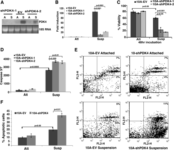
Fig 2.
Depletion of PDK4 in MCF10A cells enhances anoikis. MCF10A cells were transduced with a retroviral vector (EV) or two independent shRNAs targeting PDK4 (shPDK4) and cultured under attached (Att) and suspended (Susp) conditions for 24 h (unless otherwise indicated). Error bars represent standard deviations. (A) Northern blotting of PDK4 depletion in MCF10A cells under attached (A) and suspended (S) conditions. 18S rRNA was used as a loading control. (B) Quantitative determination of PDK4 knockdown efficiency in MCF10A cells under attached and suspended conditions. The RNA level of PDK4 in suspended MCF10A cells was set as 100. (C) Trypan blue exclusion assay of cell viability in control and PDK4-depleted MCF10A cells under attached and suspended (for 48 h) conditions. (D) Measurement of caspase 3/7 activity in control and PDK4-depleted MCF10A cells. (E) Fluorescence-activated cell sorting analysis of annexin V/7-AAD staining in control and PDK4-depleted MCF10A cells. The x axes show annexin V staining, and y axes show 7-AAD staining. (F) Statistics of total apoptotic cells based on the annexin V/7-AAD analysis.

